Protocol for Flow Cytometric Analysis of IFNAR2 and CD3 Surface Expression on Human Peripheral Blood Mononuclear Cells (PMBC)
-
Isolate peripheral blood mononuclear cells (PBMC) from heparinized venous blood obtained from healthy donors by Ficoll gradient (Type 400, Pharmacia, Uppsala, Sweden) centrifugation.
-
Collect
-
Anti-IFNAR2 MAb (PBL Catalog No. 21385-1)
-
Isotype-matched negative control MAb (mouse IgG2a, e.g. DakoCytomation (X0943).
-
Wash cells twice in NaCl. (Resuspend gently, then centrifuge at 4°C, 1800 rpm 5 min).
-
Collect 106 cells per FACS tube and wash with wash buffer.
-
Carefully pipette off the supernatant and discard.
-
Gently mix to resuspend cells in 50 μl wash buffer.
-
Block nonspecific binding sites (FcR) by adding 10 μl rabbit IgG (Sigma Aldrich, I 8140).
-
Incubate 5 min at room temperature.
-
Add the Mabs as follows:
-
Tubes 1 and 2: 3 μl negative control mouse IgG2a (DakoCytomation (X0943) final concentration 6 μg/ml)
-
Tubes 3 and 4: Anti-IFNAR2 MAb (PBL Catalog No. 21385-1) to a final concentration of 2.5 μg/ml.
-
Incubate on ice for 40 min.
-
Wash twice in wash buffer.
-
Resuspend cells in 50 μl wash buffer with biotin-conjugated goat anti-mouse IgG (Jackson ImmunoResearch (115-066-072) to a final concentration of 5 μg/ml.
-
Incubate on ice for 40 min.
-
Wash twice in wash buffer.
-
Resuspend cells in 50 μl wash buffer to include
-
Tubes 1-4: R-Phycoerythrin-conjugated Streptavidin (Jackson ImmunoResearch (016-110-084) to a final concentration of 2.5 μg/ml. In order to analyze IFNAR2 expression on T lymphocytes, further add to
-
Tubes 1 and 3: 2 μl negative control mouse IgG1-APC (DakoCytomation (X0968), final concentration 4 μg/ml).
-
Tubes 2 and 4: 1 μg/ml final concentration APC-conjugated mouse anti-human CD3 MAb (Ancell, 144-060).
-
Incubate on ice for 15 min.
-
Wash cells twice in wash buffer.
-
Resuspend cells in PBS plus 1 μg/ml propidium-iodine (to exclude dead cells from the measurements. This can be omitted).
-
Harvest cells for FACS: collect at least 50000 events contained in the live cells gate.
Image
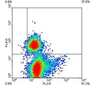
FL2-H = IFNAR2
FL4-H = CD3
Figure 1. Flow cytometric analysis of PBMC isolated from venous blood of healthy donors
and promptly stained for IFNAR2 (FL2-H) and CD3 (FL4-H) as described. The quadrant markers
were set on the bases of the fluorescence of the same PBMC stained with the corresponding
isotype-matched negative control antibodies. Numbers represent the quadrant percentiles of cells
staining positively for the indicated molecules.
Protocol for Flow Cytometric Analysis of IFNAR2 and CD3 Surface Expression on Human Peripheral Blood Mononuclear Cells (PMBC)
-
Isolate peripheral blood mononuclear cells (PBMC) from heparinized venous blood obtained from healthy donors by Ficoll gradient (Type 400, Pharmacia, Uppsala, Sweden) centrifugation.
-
Collect
-
Anti-IFNAR2 MAb (PBL Catalog No. 21385-1)
-
Isotype-matched negative control MAb (mouse IgG2a, e.g. DakoCytomation (X0943).
-
Wash cells twice in NaCl. (Resuspend gently, then centrifuge at 4°C, 1800 rpm 5 min).
-
-
Collect 106 cells per FACS tube and wash with wash buffer.
-
Carefully pipette off the supernatant and discard.
-
Gently mix to resuspend cells in 50 μl wash buffer.
-
Block nonspecific binding sites (FcR) by adding 10 μl rabbit IgG (Sigma Aldrich, I 8140).
-
Incubate 5 min at room temperature.
-
Add the Mabs as follows:
-
Tubes 1 and 2: 3 μl negative control mouse IgG2a (DakoCytomation (X0943) final concentration 6 μg/ml)
-
Tubes 3 and 4: Anti-IFNAR2 MAb (PBL Catalog No. 21385-1) to a final concentration of 2.5 μg/ml.
-
-
Incubate on ice for 40 min.
-
Wash twice in wash buffer.
-
Resuspend cells in 50 μl wash buffer with biotin-conjugated goat anti-mouse IgG (Jackson ImmunoResearch (115-066-072) to a final concentration of 5 μg/ml.
-
Incubate on ice for 40 min.
-
Wash twice in wash buffer.
-
Resuspend cells in 50 μl wash buffer to include
-
Tubes 1-4: R-Phycoerythrin-conjugated Streptavidin (Jackson ImmunoResearch (016-110-084) to a final concentration of 2.5 μg/ml. In order to analyze IFNAR2 expression on T lymphocytes, further add to
-
Tubes 1 and 3: 2 μl negative control mouse IgG1-APC (DakoCytomation (X0968), final concentration 4 μg/ml).
-
Tubes 2 and 4: 1 μg/ml final concentration APC-conjugated mouse anti-human CD3 MAb (Ancell, 144-060).
-
-
-
Incubate on ice for 15 min.
-
Wash cells twice in wash buffer.
-
Resuspend cells in PBS plus 1 μg/ml propidium-iodine (to exclude dead cells from the measurements. This can be omitted).
-
Harvest cells for FACS: collect at least 50000 events contained in the live cells gate.
Related Article
A Cost-Effective Antibody Cocktail For Broad Neutralization Of Human Type I IFN Activity
Read Article