Paradoxically, there are relatively few commercially available reagents for the study of macaque cytokines including type I interferons. Type I interferons, including interferon alpha (IFN-α), play critical roles in the host immune responses to pathogens. Conversely, aberrant expression of IFN-α has been shown to be associated with chronic inflammation and autoimmunity. Therefore, development of reagents to determine the levels of IFN-α in nonhuman primates should foster an improved understanding of this cytokine family in a highly relevant model system, allow for the development of better antiviral therapeutics, and facilitate development of treatments for autoimmune disorders such as systemic lupus erythematosus.
The detection of IFN-α proteins can be accomplished by biological activity measures or by immunodetection methods (Jubin R (2008) Eur Biopharm Rev 44:34-37). Biological activity assays represent the gold standard for cytokine detection. However, these assays are technically challenging, time consuming, and often exhibit cross-reactivity with related cytokines. Therefore, simpler, more rapid assays that can selectively distinguish analytes such as IFN-α in complex mixtures are highly desirable. PBL has recently developed a sensitive, rapid sandwich ELISA kit (PBL # 46100-1) for the determination of macaque (Rhesus and Cynomolgus) IFN-α.
Evaluation
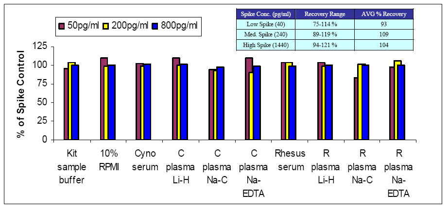
In an effort to better understand the performance of the new Cynomolgus/Rhesus IFN-α serum ELISA in a variety of sample matrices, a spike recovery study was performed. Briefly, Rhesus/Cynomolgus IFN-α2 protein (PBL # 14110-1) was spiked separately into RPMI/10% FBS tissue culture media, Cynomolgus serum or Cynomolgus plasma (prepared by three common methods, namely, lithium heparin, sodium citrate, and sodium EDTA). Spiked samples were prepared in these matrices at low (50 pg/ml), medium (200 pg/ml), and high (800 pg/ml) concentrations. As called for in the kit protocol, the kit standard curves were run in the same matrix in which the spike samples were prepared. Figure 1 shows that the Rhesus/Cynomolgus IFN-α2 protein was detectable in media, sample buffer, and all Cynomolgus matrices at all test concentrations with an accuracy within +/-20% of the nominal values. Similar results were obtained when studies were performed in rhesus serum and plasmas. This kit then exhibits good performance characteristics in a wide variety of sample matrices. To further illustrate matrix compatibility, a second study was performed to examine the spike recovery in eight separate lots of Cynomolgus serum (inset table). Similar to the initial matrix study, these results demonstrate consistent recovery using several lots of serum, suggesting that the kit should perform well when large sample sets are analyzed.
Figure 1. Spike recovery of Cyno Interferon Alpha in NHP sera and plasma matrices
using PBL's Cyno/Rhesus IFN Alpha ELISA kit (46100)
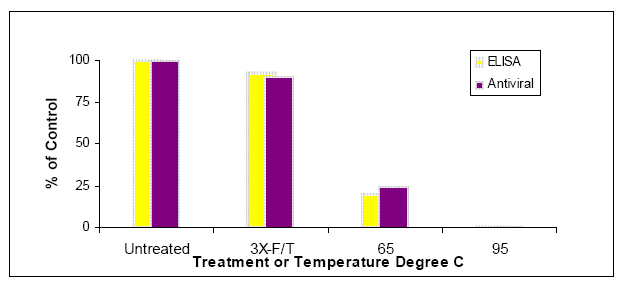
Cytokines such as IFN-α are functional proteins that require properly folded forms of the proteins to interact properly with their cognate receptors leading to modulation of gene expression. Low activity/mass ratios of the IFN in a bioassay would suggest that the sample contains a high percentage of inactive forms of proteins. Ideally, an ELISA would preferentially detect only the active forms of the IFN-α, thus providing a good correlate to the bioassay for IFN-α in experimental samples. To examine the correlation between this ELISA kit and bioactivity results, IFN-α2 samples were prepared at a concentration of 1600 pg/ml and then subjected to several stressors. After three freeze/thaw cycles, the protein showed little loss in activity in bioassay (Figure 2), and the ELISA data demonstrated similar stability of the IFN-α2 protein in response to this treatment. Heat inactivation at either 65ºC or 95ºC for 1 hr was used to denature the IFN-α2 protein. Samples lost a majority of their bioactivity in the 65ºC treatment, and exhibited a near complete loss of biological activity when heated to 95ºC. Again, the protein level measured by ELISA was similar to that expected from bioassay results suggesting that the ELISA preferentially detects the bioactive, functional form of the Rhesus/Cynomolgus IFN-α2 protein.
Figure 2.PBL's Cyno/Rhesus IFN Alpha ELISA (46100) selectively quantifies intact,
biologically active non-human primate IFN alpha protein
This brief report on the new VeriKine Cynomolgus/Rhesus IFN-α Serum ELISA kit (PBL # 46100-1) summarizes the performance of the kit in a range of sample matrices anticipated to be used by researchers. In addition, the kit preferentially detects the active form of the Rhesus/Cynomolgus IFN-α2 protein. This latter result suggests that this kit may serve as an appropriate correlate for the more complex bioassays and, therefore, may expedite research by minimizing the number of samples to be analyzed in more time-consuming bioassays.
Paradoxically, there are relatively few commercially available reagents for the study of macaque cytokines including type I interferons. Type I interferons, including interferon alpha (IFN-α), play critical roles in the host immune responses to pathogens. Conversely, aberrant expression of IFN-α has been shown to be associated with chronic inflammation and autoimmunity. Therefore, development of reagents to determine the levels of IFN-α in nonhuman primates should foster an improved understanding of this cytokine family in a highly relevant model system, allow for the development of better antiviral therapeutics, and facilitate development of treatments for autoimmune disorders such as systemic lupus erythematosus.
The detection of IFN-α proteins can be accomplished by biological activity measures or by immunodetection methods (Jubin R (2008) Eur Biopharm Rev 44:34-37). Biological activity assays represent the gold standard for cytokine detection. However, these assays are technically challenging, time consuming, and often exhibit cross-reactivity with related cytokines. Therefore, simpler, more rapid assays that can selectively distinguish analytes such as IFN-α in complex mixtures are highly desirable. PBL has recently developed a sensitive, rapid sandwich ELISA kit (PBL # 46100-1) for the determination of macaque (Rhesus and Cynomolgus) IFN-α.
Evaluation
In an effort to better understand the performance of the new Cynomolgus/Rhesus IFN-α serum ELISA in a variety of sample matrices, a spike recovery study was performed. Briefly, Rhesus/Cynomolgus IFN-α2 protein (PBL # 14110-1) was spiked separately into RPMI/10% FBS tissue culture media, Cynomolgus serum or Cynomolgus plasma (prepared by three common methods, namely, lithium heparin, sodium citrate, and sodium EDTA). Spiked samples were prepared in these matrices at low (50 pg/ml), medium (200 pg/ml), and high (800 pg/ml) concentrations. As called for in the kit protocol, the kit standard curves were run in the same matrix in which the spike samples were prepared. Figure 1 shows that the Rhesus/Cynomolgus IFN-α2 protein was detectable in media, sample buffer, and all Cynomolgus matrices at all test concentrations with an accuracy within +/-20% of the nominal values. Similar results were obtained when studies were performed in rhesus serum and plasmas. This kit then exhibits good performance characteristics in a wide variety of sample matrices. To further illustrate matrix compatibility, a second study was performed to examine the spike recovery in eight separate lots of Cynomolgus serum (inset table). Similar to the initial matrix study, these results demonstrate consistent recovery using several lots of serum, suggesting that the kit should perform well when large sample sets are analyzed.
Cytokines such as IFN-α are functional proteins that require properly folded forms of the proteins to interact properly with their cognate receptors leading to modulation of gene expression. Low activity/mass ratios of the IFN in a bioassay would suggest that the sample contains a high percentage of inactive forms of proteins. Ideally, an ELISA would preferentially detect only the active forms of the IFN-α, thus providing a good correlate to the bioassay for IFN-α in experimental samples. To examine the correlation between this ELISA kit and bioactivity results, IFN-α2 samples were prepared at a concentration of 1600 pg/ml and then subjected to several stressors. After three freeze/thaw cycles, the protein showed little loss in activity in bioassay (Figure 2), and the ELISA data demonstrated similar stability of the IFN-α2 protein in response to this treatment. Heat inactivation at either 65ºC or 95ºC for 1 hr was used to denature the IFN-α2 protein. Samples lost a majority of their bioactivity in the 65ºC treatment, and exhibited a near complete loss of biological activity when heated to 95ºC. Again, the protein level measured by ELISA was similar to that expected from bioassay results suggesting that the ELISA preferentially detects the bioactive, functional form of the Rhesus/Cynomolgus IFN-α2 protein.
This brief report on the new VeriKine Cynomolgus/Rhesus IFN-α Serum ELISA kit (PBL # 46100-1) summarizes the performance of the kit in a range of sample matrices anticipated to be used by researchers. In addition, the kit preferentially detects the active form of the Rhesus/Cynomolgus IFN-α2 protein. This latter result suggests that this kit may serve as an appropriate correlate for the more complex bioassays and, therefore, may expedite research by minimizing the number of samples to be analyzed in more time-consuming bioassays.