Protocol for Flow Cytometric Analysis of IFNAR2 and CD3 Surface Expression on Human Peripheral Blood Mononuclear Cells (PMBC)
- Isolate peripheral blood mononuclear cells (PBMC) from heparinized venous blood obtained from healthy donors by Ficoll gradient (Type 400, Pharmacia, Uppsala, Sweden) centrifugation.
- Collect
- Anti-IFNAR2 MAb (PBL Catalog No. 21385-1)
- Isotype-matched negative control MAb (mouse IgG2a, e.g. DakoCytomation (X0943).
- Wash cells twice in NaCl. (Resuspend gently, then centrifuge at 4°C, 1800 rpm 5 min).
- Collect 106 cells per FACS tube and wash with wash buffer.
- Carefully pipette off the supernatant and discard.
- Gently mix to resuspend cells in 50 μl wash buffer.
- Block nonspecific binding sites (FcR) by adding 10 μl rabbit IgG (Sigma Aldrich, I 8140).
- Incubate 5 min at room temperature.
- Add the Mabs as follows:
- Tubes 1 and 2: 3 μl negative control mouse IgG2a (DakoCytomation (X0943) final concentration 6 μg/ml)
- Tubes 3 and 4: Anti-IFNAR2 MAb (PBL Catalog No. 21385-1) to a final concentration of 2.5 μg/ml.
- Incubate on ice for 40 min.
- Wash twice in wash buffer.
- Resuspend cells in 50 μl wash buffer with biotin-conjugated goat anti-mouse IgG (Jackson ImmunoResearch (115-066-072) to a final concentration of 5 μg/ml.
- Incubate on ice for 40 min.
- Wash twice in wash buffer.
- Resuspend cells in 50 μl wash buffer to include
- Tubes 1-4: R-Phycoerythrin-conjugated Streptavidin (Jackson ImmunoResearch (016-110-084) to a final concentration of 2.5 μg/ml. In order to analyze IFNAR2 expression on T lymphocytes, further add to
- Tubes 1 and 3: 2 μl negative control mouse IgG1-APC (DakoCytomation (X0968), final concentration 4 μg/ml).
- Tubes 2 and 4: 1 μg/ml final concentration APC-conjugated mouse anti-human CD3 MAb (Ancell, 144-060).
- Tubes 1-4: R-Phycoerythrin-conjugated Streptavidin (Jackson ImmunoResearch (016-110-084) to a final concentration of 2.5 μg/ml. In order to analyze IFNAR2 expression on T lymphocytes, further add to
- Incubate on ice for 15 min.
- Wash cells twice in wash buffer.
- Resuspend cells in PBS plus 1 μg/ml propidium-iodine (to exclude dead cells from the measurements. This can be omitted).
- Harvest cells for FACS: collect at least 50000 events contained in the live cells gate.
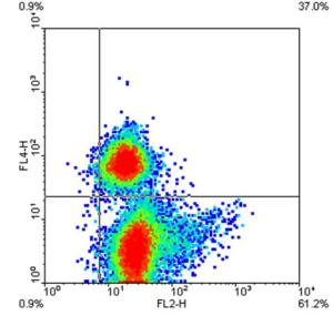
FL2-H = IFNAR2
FL4-H = CD3
Figure 1. Flow cytometric analysis of PBMC isolated from venous blood of healthy donors
and promptly stained for IFNAR2 (FL2-H) and CD3 (FL4-H) as described. The quadrant markers
were set on the bases of the fluorescence of the same PBMC stained with the corresponding
isotype-matched negative control antibodies. Numbers represent the quadrant percentiles of cells
staining positively for the indicated molecules.